Cisterns of brain and its contents along with its classification and
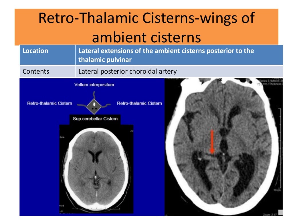
The ambient cistern is a thin, sheet-like extension of the quadrigeminal cistern that extends laterally around the midbrain and posterior to the thalami. It acts as the connection between the quadrigeminal cistern and the interpeduncular cistern.
Cureus Delineation of Subarachnoid Cisterns Using CT Cisternography, CT Brain Positive and
The anterior circulation of the brain is related to the subarachnoid cisterns of the supratentorial region and the cisternal approach to the supratentorial lesions consists of a compartmental opening of one or more of these cisterns (88, 97, 98, 100, 105-107). Any lesion may grow epiarachnoidally or also within one or more cisterns but, in both.
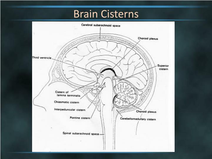
PPT Brain Cisterns PowerPoint Presentation ID2263744
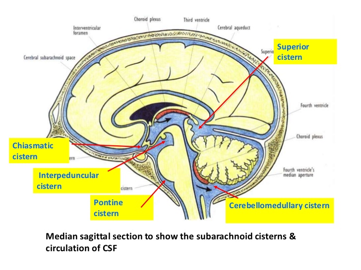
The prepontine cistern, or simply pontine cistern , is an unpaired CSF-filled subarachnoid cistern located ventral to the pons and dorsal to the clivus. It is bounded by arachnoid membranes which separate it from surrounding cisterns. superiorly the mesencephalic leaf of the membrane of Liliequist, above which is the interpeduncular cistern.

MRI Brain ambiens cistern anatomy Radiology Anatomy Images Mri brain, Mri, Brain anatomy
0:00 / 15:04 Ventricles and Cisterns of the Brain | Radiology anatomy part 1 prep | MRI brain Radiology Tutorials 37.3K subscribers Subscribe 715 20K views 1 year ago Anatomy Tutorials High.
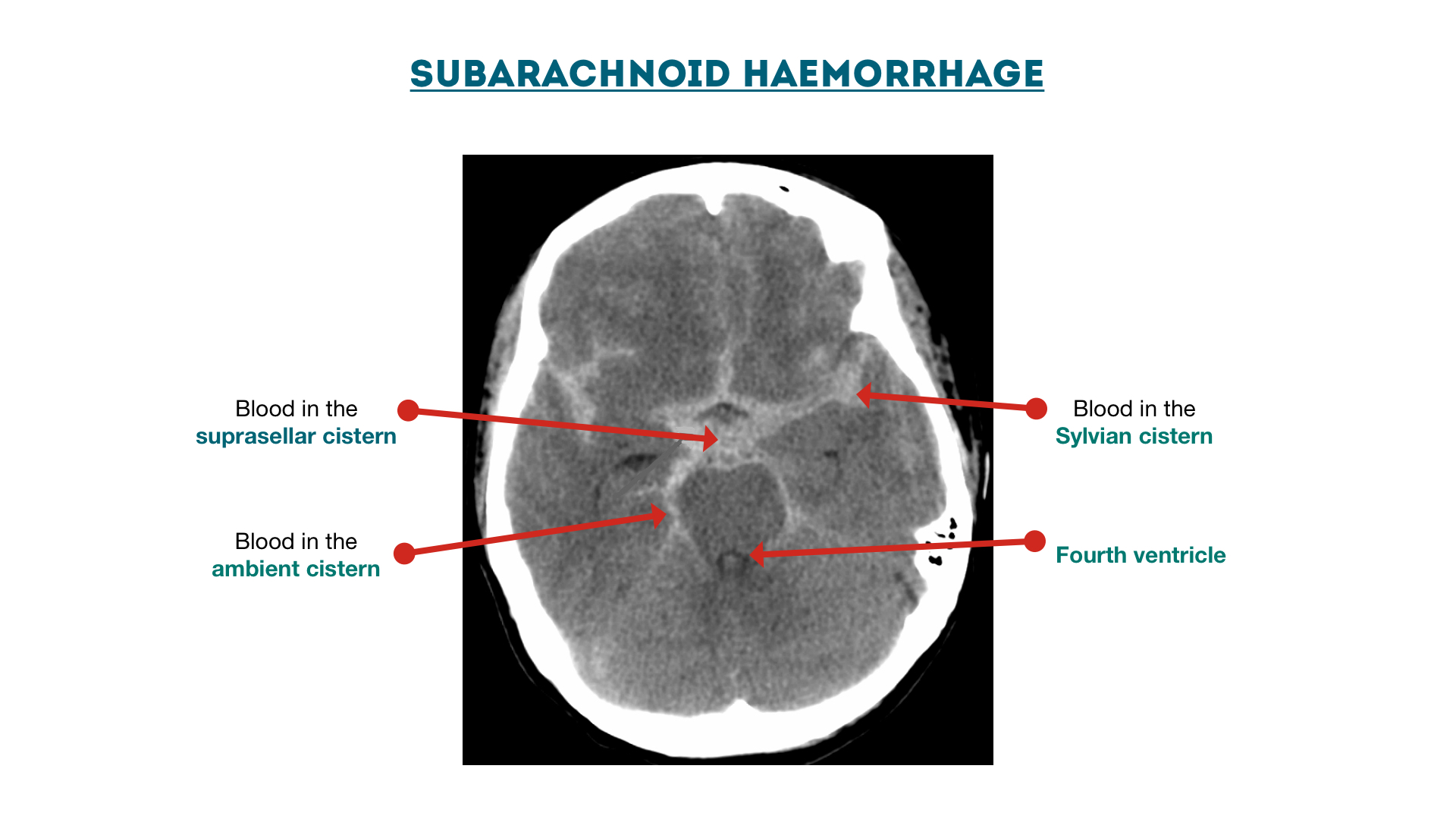
Subarachnoid haemorrhage cisterns Geeky Medics
Subarachnoid cisterns - bathes the brain, between arachnoid mater and pia mater. Here the CSF is reabsorbed back into the circulation. By TeachMeSeries Ltd (2024) Fig 3 - The anatomical positioning of the ventricles of the brain. Production and Reabsorption of Cerebrospinal Fluid.

PPT Brain Cisterns PowerPoint Presentation ID2263744
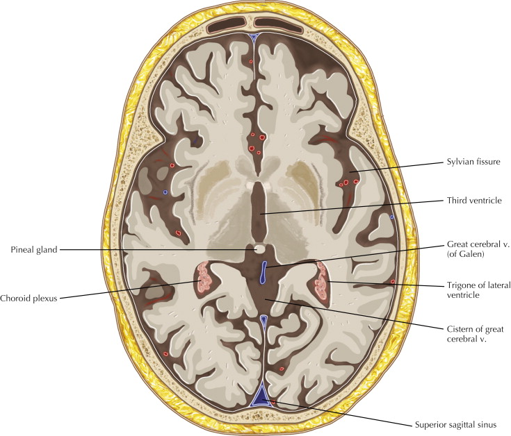
The subarachnoid cisterns are clinically and surgically significant enlarged cerebrospinal fluid (CSF)-filled pockets of the subarachnoid space that transmit important neurovascular structures including cranial nerves and intracranial vessels.
ventricular system overview Brain Imaging
The interpeduncular cistern is located at the base of the brain at the junction where the arachnoid mater stretches between the two temporal lobes, occupying the interpeduncular fossa. Located within this cistern is the optic chiasm as well as some important neurovascular structures which include:.
Basal CSF cisternsInterpeduncular cistern RANZCRPart1 Wiki FANDOM powered by Wikia
The interpeduncular cistern is an unpaired CSF-filled subarachnoid cistern located between the cerebral peduncles. It is partially bounded by the leaves of the Liliequist membrane, one of the arachnoid membranes , which separate it from its direct cranial and caudal relations 1.

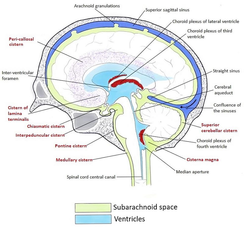
Normal sagittal midline MRI showing the CSF cisterns. A. Cistern of the laminae terminalis. B
The subarachnoid cisterns are spaces formed by openings in the subarachnoid space, an anatomic space in the meninges of the brain. [1] The space is situated between the two meninges, the arachnoid mater and the pia mater. These cisterns are filled with cerebrospinal fluid (CSF). [1] Structure

suprasellar cistern anatomy
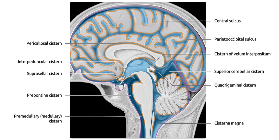
The subarachnoid space is described as a cistern at points where spaces exist between it and the underlying pia mater. At different points around the brain, the cisterns are described with respect to adjacent anatomical landmarks. Notable cisterns include the: suprasellar or chiasmatic cistern; interpeduncular cistern; prepontine cistern

6 Basal Cisterns Anatomy Neupsy Key
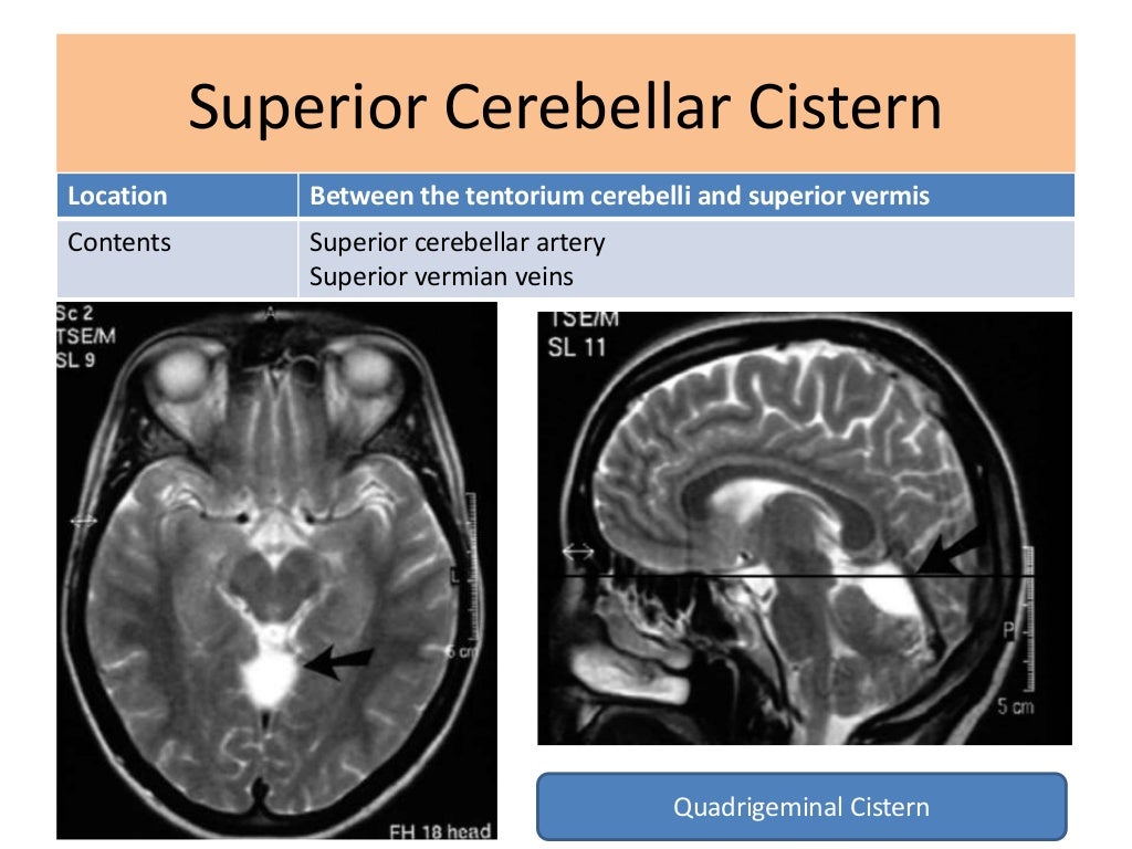
Ambient cistern (Figure 6): Located in the lateral aspect of the brain stem. This cistern has a particular surgical importance as it has both supratentorial and infratentorial extensions. Divided by the superior cerebellar membrane, the superior compartment or posterior cerebral ambient cistern houses the posterior cerebral artery, medial and.
Subarachnoid cisterns Anatomy and clinical points Kenhub
Normal Anatomy The ventricular system is located deep within the brain and is filled with cerebrospinal fluid (CSF). The normal appearance of CSF on magnetic resonance images is that of water. This axial MR image shows the choroid plexus within the ventricles, where CSF is produced.
Ventricles and Cerebrospinal Fluid Cisterns Radiology Key
The subarachnoid cisterns, or basal cisterns , are compartments within the subarachnoid space where the pia mater and arachnoid membrane are not in close approximation and cerebrospinal fluid (CSF) forms pools or cisterns (Latin: "box"). As they are interconnected, their patency is essential for CSF circulation.

2 Ventricles and Cisterns Radiology Key
The concept of intracranial surgery in terms of moving from one cistern to another is presented here with particular emphasis on the cisterns in surgical approaches to intracranial vessels and nerves for the treatment of aneurysms, arteriovenous malformations, and for surgery of basal tumors. MeSH terms Brain / blood supply

PPT Brain Cisterns PowerPoint Presentation ID2263744
A cisternogram scan is a diagnostic procedure that evaluates how cerebrospinal fluid (CSF) flows around your brain and spinal cord. This protective fluid: Delivers nutrients to your brain and spine. Helps your central nervous system (CNS) work. Removes toxins from tissues in your CNS. Acts like a cushion to protect your brain from concussion.
Cisterns of brain and its contents along with its classification and
Definition and location of the falx cerebri. The brain is bathed in fluid during life. The name of this substance is cerebrospinal fluid (CSF). It provides the brain with nutrients, allows for solute exchange, and provides basic mechanical and functional support to the organ.
